Description
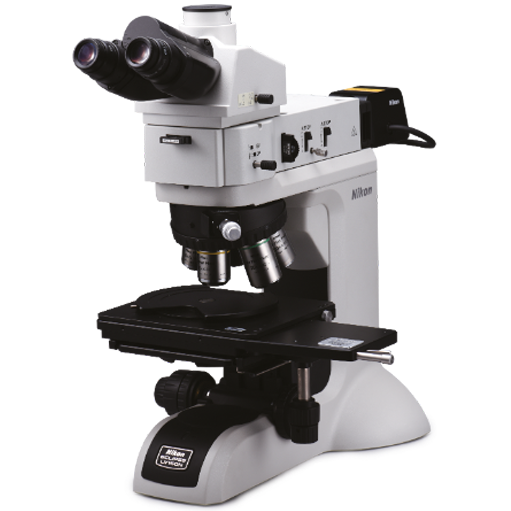
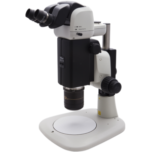
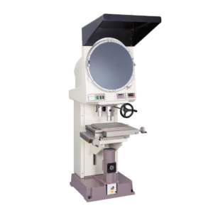
The Nikon LV150N wafer inspection microscope is suitable for wafers from 4″-6″. The mounting plate can be rotated and the cross table is equipped with quick adjustment. The high-quality Nikon lenses guarantee bright, high-contrast, clear and flat images at incredible working distances with high resolution.
MMK-Snap 5 is a powerful complete package consisting of microscope camera, USB cable, dongle, measuring scale and PC software
“Recordings – measurements – generation of depth-of-field images”
The clear and uncluttered operation of the software enables every user to take images and carry out measurements in no time at all! The all-in-one set is therefore ideal for use in the industrial sector, but also in a university or institutional environment, especially when many people want to get results quickly and perhaps only work on the system from time to time. A particular highlight is the ability to combine images with different depth of field ranges directly into a completely sharp resulting image.
Powerful microscope camera and microscope software. Made in Germany!
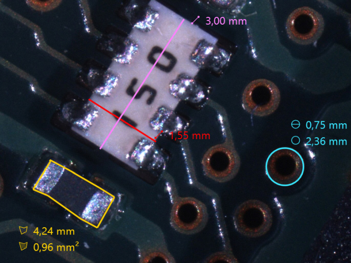
Measuring functions:
- Distance measurement (single and polygonal)
- Circular measurement (radius, diameter, area, circumference)
- Area measurement (area, perimeter)
- Angle measurement
- Perpendicular measurement
- Counting function
- Fade-in scale bar
![]()
![]()
![]()
![]()
![]()
![]()
![]()
![]()
Camera functions:
- Exposure time can be set once, continuously or manually
- White balance directly adjustable at the touch of a button
- Signal amplification can be set once, continuously or manually
- Color saturation manually adjustable
- Live image can be mirrored horizontally / vertically
- Digital resharpening function
- Shading
- Fade-in crosshairs
- Depth of field function
- Panorama function
![]()
![]()
![]()
![]()
![]()
![]()
![]()
![]()
![]()
![]()
![]()
Labeling and auxiliary functions:
- Marking arrow
- Text field
- Guideline
![]()
![]()
![]()
General functions:
- Open image
- Storage function in JPG, BMP, PNG and TIF
- Interface to the Windows clipboard
- Direct print function
- Setting options for font, color and line thickness
![]()
![]()
![]()
![]()
![]()
![]()
![]()
Camera data
- Resolution 5 mega pixels (2448 x 2048 pixels)
- 2/3″ Sony IMX264 CMOS sensor color, global shutter
- Frame rate: max. 38 fps
- USB 3.1 connection
- C-mount
Supported languages
The language for operating the software can be changed directly in the help menu if required (instructions in German and English).
- German
- English
- French
- Italian
- Polish
- Portuguese
- Romanian
- Spanish
- Czech
- Hungarian
Scope of delivery:
- Microscope camera 5 Mpix, 2/3″
- USB 3.1 connection cable type A
- Recording and measuring software (WIN10/WIN11)
- USB 3.1 license dongle type A
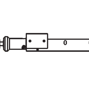
- Measuring scale
When developing the software for MMK-Snap 5, the main focus was on very simple and intuitive operation. From installation, image acquisition and measurement to exporting the analyzed image, every user can complete the image acquisition and measurement within a very short time. After starting the software, the live image opens directly and can be started.
A particular highlight is the ability to combine images with different depth of field ranges directly into a completely sharp resulting image. All you have to do is start the recording and press the focus drive on the microscope -> the resulting image is created immediately after the recording stops. Once the image has been captured, you can switch directly to the measurement area. The settings for annotations and measurements, such as color, thickness and font size, can be changed quickly via an intuitive right-click menu. The image can now be saved to any location in the file system using a save button, or copied directly to the clipboard and pasted from there into a report, for example.
Note: This is a general description and the illustrations are examples.

