


Description
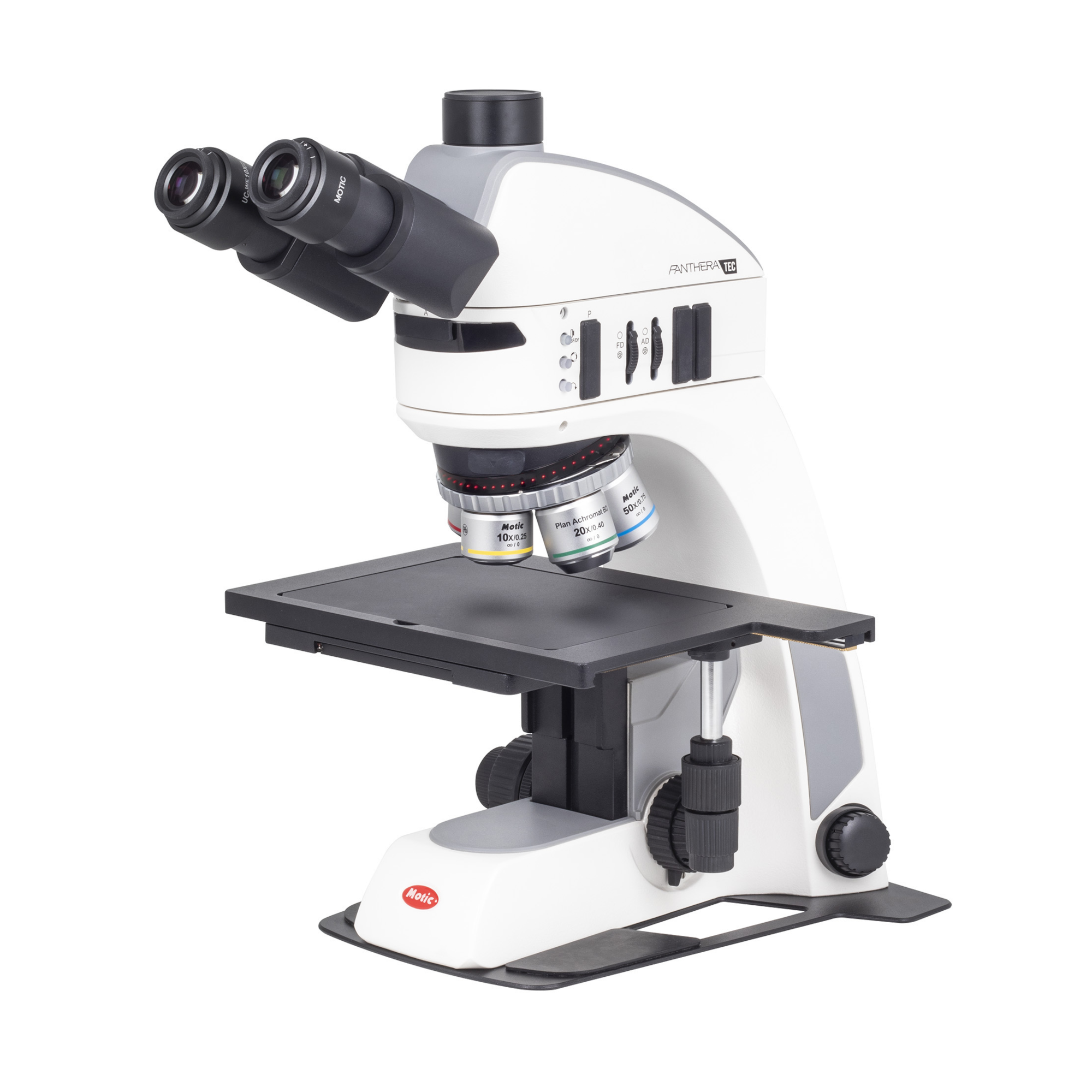
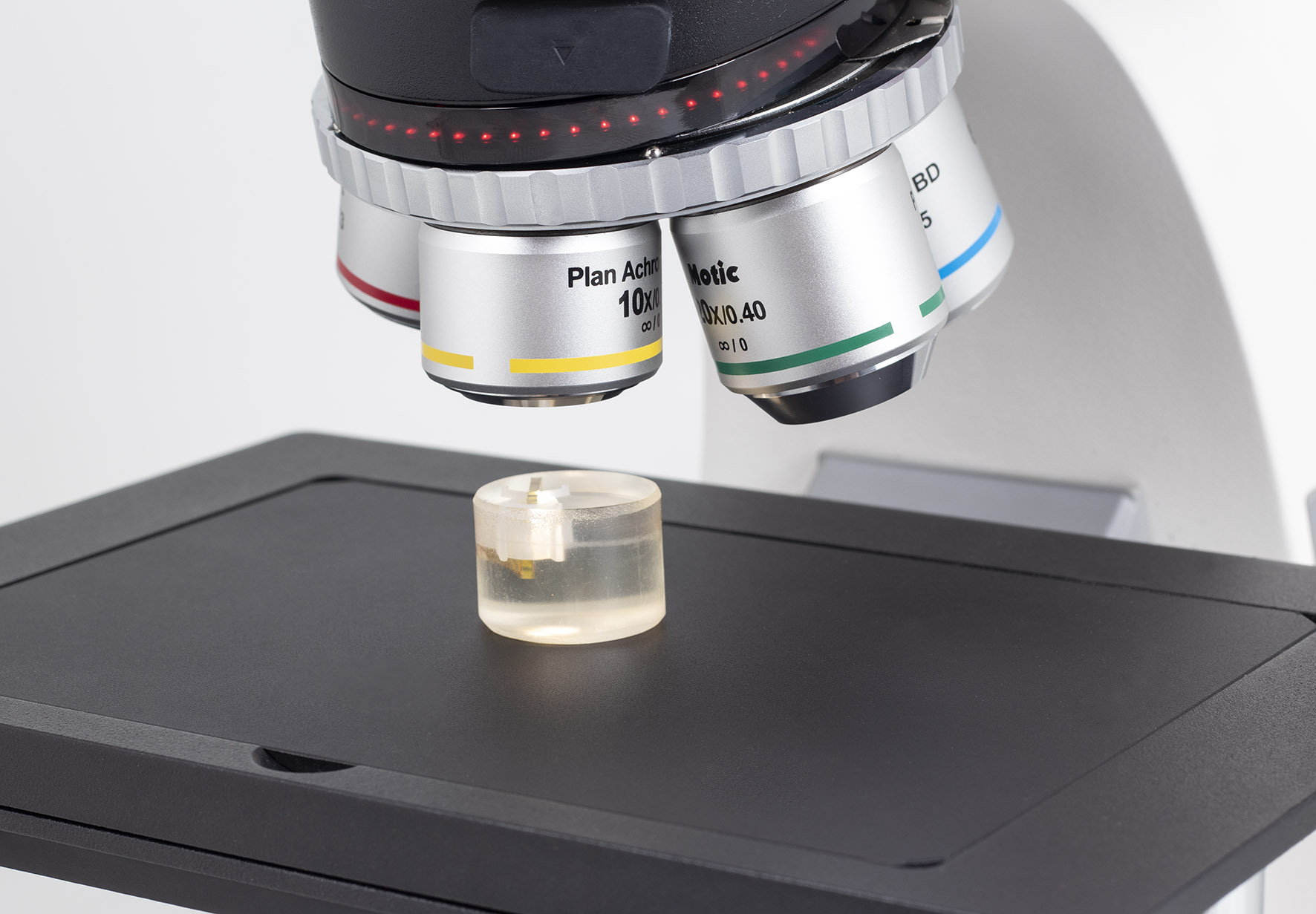
The Panthera TEC models offer a wide range of possibilities for materials science, particularly in technical training and industrial quality assurance. Brightfield, darkfield, and simple polarization contrast are achieved through a new LED illumination concept. The 5-segment ring light enables darkfield and oblique incident illumination—perfect for detecting scratches or other defects on flat, reflective surfaces without tilting the specimen.
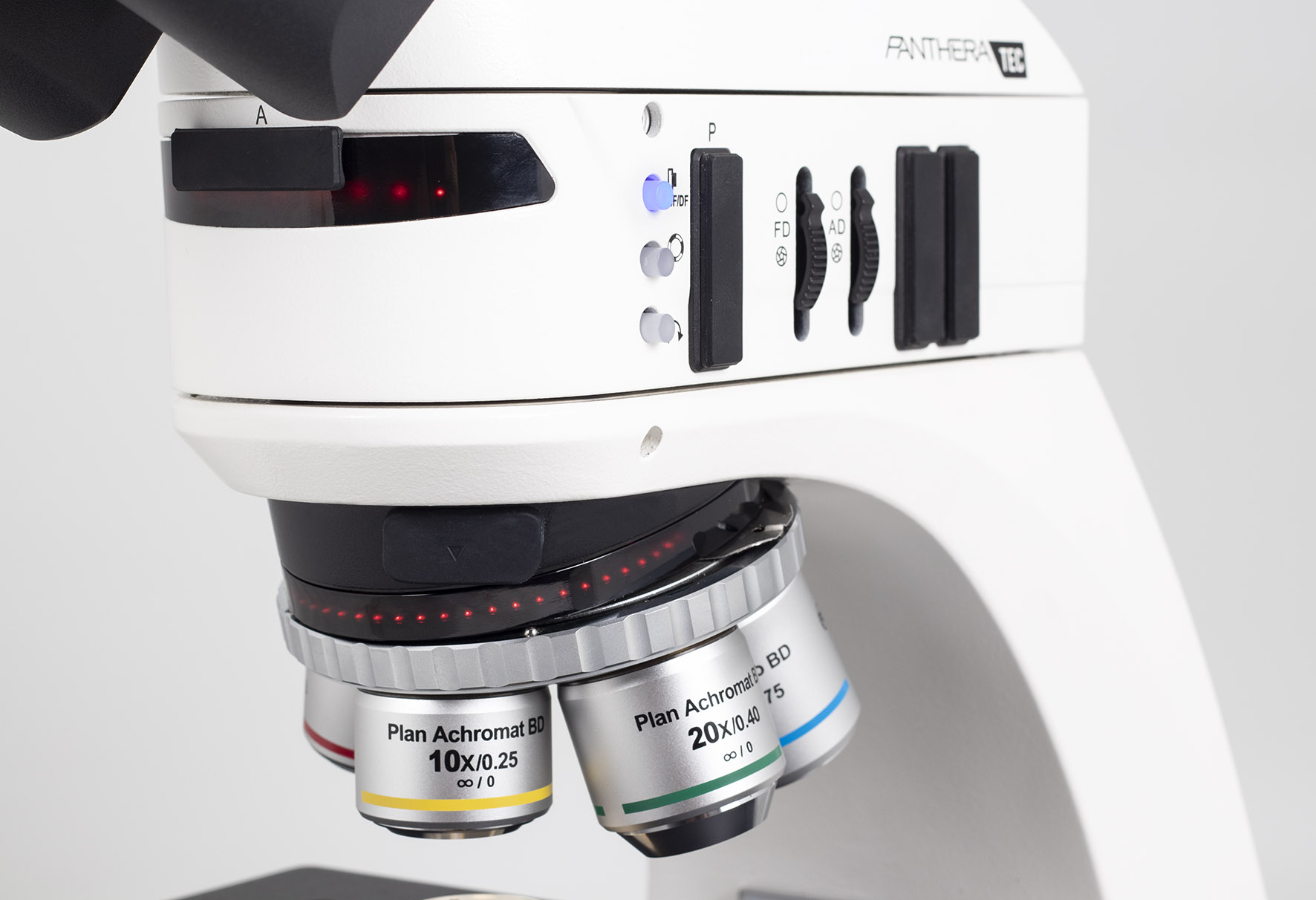
The Panthera TEC BD models feature a coded 5-position nosepiece equipped with LD Plan HF/DF objectives that deliver excellent image quality. The coded nosepiece automatically saves the last brightness setting for each position and restores it as soon as the objective is rotated back into place.
The compact incident-light illuminator includes slots for a polarizer and analyzer, enabling simple polarization contrast while also preventing image-degrading internal reflections. The illumination mode (HF/DF) and the illumination angle (via segment control) are adjusted directly on the illuminator. The beam split on the trinocular models (25° viewing angle) is fixed at 50:50 (visual/camera output).
The Panthera TEC models are clearly designed for applications in materials science.
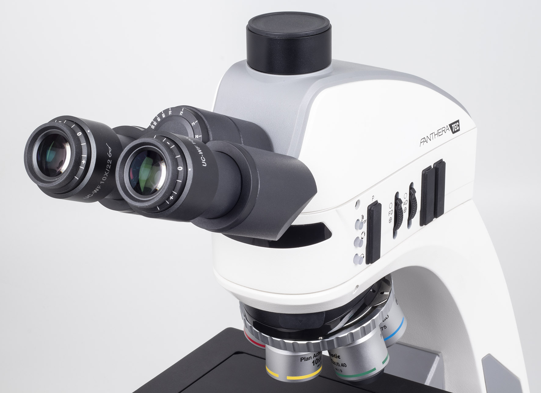
Trinocular head reversed with 25° viewing angle
Fixed 50:50 beam split (eyepiece:camera)
Interpupillary distance 48–75 mm
10x eyepieces with diopter adjustment
LED reflected-light illuminator with field and aperture diaphragms
Polarizer and analyzer
5-position reversed nosepiece, coded with Motic LightTracer (Intelligent Light Management)
Objectives: 5X/0.13 (WD 20.3 mm), 10X/0.25 (WD 17.5 mm), 20X/0.40 (WD 8.1 mm), S-Apo 50X/0.80 (WD 1 mm)
Mechanical X/Y stage
Stage size 300 x 180 mm
XY travel range 150 x 100 mm (6″ x 4″)
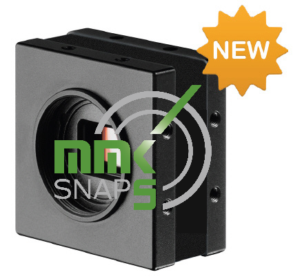
MMK-Snap 5 is a powerful complete package consisting of microscope camera, USB cable, dongle, measuring scale and PC software
“Recordings – measurements – generation of depth-of-field images”
The clear and uncluttered operation of the software enables every user to take images and carry out measurements in no time at all! The all-in-one set is therefore ideal for use in the industrial sector, but also in a university or institutional environment, especially when many people want to get results quickly and perhaps only work on the system from time to time. A particular highlight is the ability to combine images with different depth of field ranges directly into a completely sharp resulting image.
Powerful microscope camera and microscope software. Made in Germany!
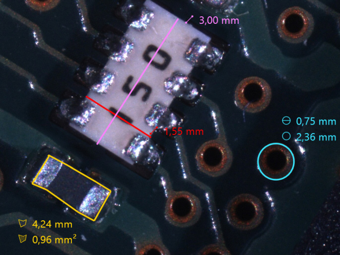
Measuring functions:
- Distance measurement (single and polygonal)
- Circular measurement (radius, diameter, area, circumference)
- Area measurement (area, perimeter)
- Angle measurement
- Perpendicular measurement
- Counting function
- Fade-in scale bar


Camera functions:
- Exposure time can be set once, continuously or manually
- White balance directly adjustable at the touch of a button
- Signal amplification can be set once, continuously or manually
- Color saturation manually adjustable
- Live image can be mirrored horizontally / vertically
- Digital resharpening function
- Shading
- Fade-in crosshairs
- Depth of field function
- Panorama function


Labeling and auxiliary functions:
- Marking arrow
- Text field
- Guideline


General functions:
- Open image
- Storage function in JPG, BMP, PNG and TIF
- Interface to the Windows clipboard
- Direct print function
- Setting options for font, color and line thickness


Camera data
- Resolution 5 mega pixels (2448 x 2048 pixels)
- 2/3″ Sony IMX264 CMOS sensor color, global shutter
- Frame rate: max. 38 fps
- USB 3.1 connection
- C-mount
Supported languages
The language for operating the software can be changed directly in the help menu if required (instructions in German and English).
- German
- English
- French
- Italian
- Polish
- Portuguese
- Romanian
- Spanish
- Czech
- Hungarian
Scope of delivery:
- Microscope camera 5 Mpix, 2/3″
- USB 3.1 connection cable type A
- Recording and measuring software (WIN10/WIN11)
- USB 3.1 license dongle type A
- Measuring scale
When developing the software for MMK-Snap 5, the main focus was on very simple and intuitive operation. From installation, image capture and measurement to exporting the evaluated image, every user can complete the image capture and measurement in no time at all. After starting the software, the live image opens directly and can be started.
A particular highlight is the ability to combine images with different depth of field ranges directly into a completely sharp resulting image. All you have to do is start the recording and press the focus drive on the microscope -> the resulting image is created immediately after the recording stops. Once the image has been captured, you can switch directly to the measurement area. The settings for annotations and measurements, such as color, thickness and font size, can be changed quickly via an intuitive right-click menu. The image can now be saved to any location in the file system using a save button, or copied directly to the clipboard and pasted from there into a report, for example.
Note: This is a general description and the illustrations are examples.